INTRODUCTION
The number of herniated lumbar disc (HLD) in people under 25 years of age, 5.5 per 100,000, is relatively lower than 128.3 per 100,000 for people of 40 to 45 years of age; but the frequency of the condition is increasing due to the change in body position, lifestyle, and diagnostic techniques19,20,22,23). Compared to the older people, HLD in young people tends to have soft intervertebral disc protrusion, relation to external trauma. It may have apophyseal ring fracture with similar symptoms or accompany with the lumbar disc herniation, and less severe degenerative change3). In addition, it frequently induces lower radicular pain caused by the single radiculopathy, and the neurological symptoms such as muscle weakness and dysesthesia are not as severe as older patients but severely limited by straight leg raising test. As younger patients have been reported to show better result with surgery due to the soft intervertebral disc protrusion and less severe degenerative change, surgery to younger patients including adolescents are more frequently observed when they do not respond well to conservative therapy.
The discectomy through laminectomy, which was considered traditional method, showed relatively satisfactory result, and could be applied to patients with degenerative bony spur, far lateral disc herniation, migrated and sequestrated disc. However, the surgery requires avulsion of the lumbar spine muscles, elimination of ligamentum flavum, incision of annulus fibrosus and posterior longitudinal ligament. The surgery can be complicated with postoperative damage on soft tissue, joint, nervous tissue, nerve root compression, adhesion due to scar tissue, and hemorrhage due to blood vessel damage17).
To avoid such complications of this surgery, various minimally invasive surgeries were developed1,2,10-13,15). Improvements on endoscopy, surgical instruments and surgical techniques allowed to expand the indications of endoscopy. Previous endoscopic method was to enter posterior laterally through triangular working zone to reach the center of intervertebral disc and create cavity by removing nucleus pulposus which decreases pressure in the intervertebral disc and improves therapeutic effect by removing protruded disc; however this method was limited for patients with immensely protruded disc such as nucleus pulposus herniation through posterior longitudinal ligament, and sequestrated or migrated disc1,2,4,6-14,16-18,21,24).
Recently, to overcome such limitations, extreme lateral endoscopic lumbar discectomy (ELELD) has been performed. In this study, result of ELELD based on the direction of disc protrusion, degree of protrusion and migration of the protrude disc in young patients were assessed. Additionally this study identifies indications, limitations and shortcomings of endoscopy and provides solutions.
MATERIALS AND METHODS
1. Subject
One hundred thirty-five patients out of 156 who were diagnosed with HLD underwent ELELD between December of 2007 and September of 2014 in Bundang Medical Center. All patients were male, with average age of 22 (20-25 years old). Average follow up period were 16 months. Magnetic resonance imaging (MRI) and computed tomography (CT) were done to all the patients. Surgery was done on patients who had conservative therapy for at least 3 months without improvement on lower back pain, radiating pain and radiological findings matched with clinical symptoms. Additionally, plain radiography was taken to check if iliac crest is higher than the space between L5 and S1 and enlarged transverse process prevents the working channel to get inserted to spinal epidural space. We excluded the patient who underwent laminectomy, discectomy, or endoscopic transforaminal lumbar discectomy.
The herniation was categorized as central, postero-lateral and foraminal based on the radiological findings, whether the herniated disc was migrated or not; and if migrated, severity was measured based on the posterior disc space; and if not migrated, severity was measured based on the 50% of area of lumbar spinal canal.
2. Method
The surgery was done on prone position with slight flexion and local anesthesia at the site of the herniation. C-arm was placed to observe anterior-posterior plane perpendicular to lateral plane of the upper and lower vertebrates of the target disc. Basal line was set with posterior facet line (Fig. 1) and incision was made at the baseline or 1.5 cm above the baseline as incision below the baseline gives greater chance of abdominal perforation, which as 13 to 18 cm away from the central line based on the patient’s circumstance of lumbar region and height of iliac crest. Surgery on the herniated disc between L5 and S1, extreme lateral approach could be limited due to the iliac crest and the transverse process; thus 9 to 11cm from the central line was the optimal position.
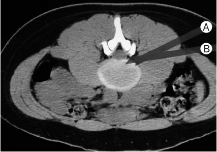
Skin incision (0.5-1 cm) was made to the injection site after local anesthesia was applied. 18 gauge needle was injected with c-arm image to inject the needle to the posterior facet by extreme lateral approach. The angle of approach was 5 to 20 degrees to the ground (Fig. 2). During the initial injection, the angle of the needle was toward the abdomen to minimize peritoneum perforation even if the needle was touching peritoneum, and changed the angle upwards to ease the access to the vertebral space at the facet joint. The position of the needle should be at the inner pedicular line from the anterior posterior view and at the intervertebral foramen from the lateral view (Fig. 3). Omnipaque 2mL was injected to the epidural space to identify relative position of the needle from the exiting nerve root and the traversing nerve root from the anterior posterior view and from the lateral view. The needle is replaced by a guide wire followed by an obturator and a final working cannula in that sequence (Fig. 4). Using the slanted working channel, the wider surgical view was provided and helped preventing nerve root damage. When the working channel passed through the exiting nerve root, slanted side was turned toward the nerve root. After that, slanted side was turned 180 degrees making the slanted side to face the traversing nerve root to minimize the damage. When endoscope with two tubing system with 6mm inner diameter and 20 degrees slope (Yeung Endoscopic Spine Surgery System [YESS]) was inserted to epidural space, blue colored lesion could be identified. During the surgery, saline mixed with antibiotics was used continuously to prevent the infection and the damage from burn. When transforaminal ligament was obstructing, the ligament was removed, and identified the colored protruding nucleus populous and removed with pituitary forcep until the nerve was decompressed and performed hemostasis using radiofrequency trigger-flex bipolar. When the HLD was removed and the decompression of the traversing nerve root was observed via endoscope, the surgery was finished. Patients were allowed to communicate and when the patient complained about symptoms related to irritating nerve root or any other neurological symptoms, the surgery were stopped immediately and continued after the problem was fixed. When epidural hemorrhage occurred during the surgery, radiofrequency trigger-flex bipolar was used to stop the bleeding and if not corrected, pressure was applied with epinephrine soaked gel foam to the hemorrhagic site and increased the tubing pressure to wash with saline. There were no serious complications including damaged nerve root, internal organ or massive hemorrhage.
3. Statistical Analysis
The postoperative follow up study was carried out by means of outpatient clinic and telephone interview, the clinical result was evaluated through MacNab’s criteria and visual analogue scale where the ‘excellent’ and ‘good’ score was considered as successful result. The statistical testing was carried out by analysis of variance (ANOVA) using SPSS for windows (Release 7.5.1; SPSS Inc., Chicago, IL, USA).
RESULTS
1. Clinical Characteristics
Sixty seven percent of patients complained about lower back pain and radicular pain, and 33% only complained about the radicular pain. Sixty five point seven percent of patients had sensory dysfunction and motor weakness was observed in 40.3%. Eighty seven percent were positive in the straight leg raising test. Patents with osteophyte due to the apophyseal ring fracture were excluded from this study. Average period of lower back pain until surgery was 2.3 years and 4.5 months for radicular pain. Seventy four point one percent of patients had surgery between L4 and L5, 19.3% had surgery on the disc between L5 and the S1, and 6.7% had surgery on the disc between L3 and L4 (Table 1). The direction of herniation was categorized into central, posterolateral and foraminal herniation and presented at 24.4%, 63% and 12.6% respectively. There was no patient with extreme lateral lumbar herniation (Table 2). In the group of patients without migration, 17.6% showed spinal canal compromise more than 50%. Twenty point six point showed mild migrated disc under the height of the posterior lumbar disc, and 6.6% showed severe migrated disc (Table 3).
2. Result of Surgery
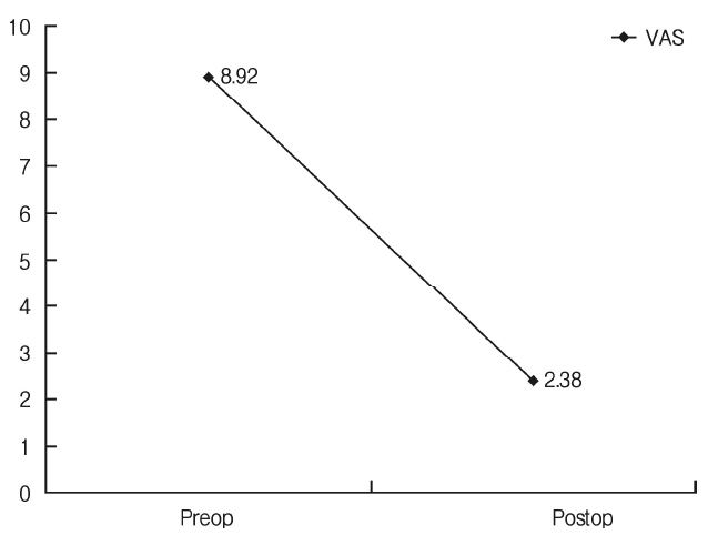
The surgery was evaluated using MacNab method and excellent or good scores were considered as successful surgery. Overall, the average visual analogue scale (VAS) of all patients before surgery was 8.92±1.20 and VAS at the final follow up was 2.38±1.55 (p<0.05) (Fig. 5). Group without migrated disc with 50% or more protrudes disc and group of mid-severe vertical migrated disc showed significant decrease in the success rate of the surgery (p<0.05) (Table 4). Post-operative complications included dysesthesia, discitis, hemorrhage, cerebrospinal fluid leakage, decrease of lumbar disc, and nerve root damage (Table 5). Twenty percent of the patients showed dysesthesia after surgery, but most patients except for 4 recovered after 3 to 4 weeks, and 2 patients had spinal fluid leak, but these were corrected with conservatory therapy such as bed rest. Discitis and decrease in the height of the lumbar disc were observed with one patient, but discitis was treated with antibiotics and only conservative therapy. Therefore, posterior lumbar interbody fusion was not performed because of lack of severe lower back pain. Two patients had severe lower back pain due to the decrease in lumbar disc height after surgery, but corrected with posterior lumbar interbody fusion. Out of 8 patients who had ‘fair’ result, 2 had multiple herniations, 3 had lasting dysesthesia after surgery, and 3 had incomplete removal of nucleus pulposus. Patients with ‘fair’ result had chiropractic therapy after surgery and reoperation was not performed. Out of 6 patients with bad result, one patient presented with decrease of disc space due to discitis who had mild herniation without disc migration, 2 patients received posterior interbody fusion as they presented with severe lower back pain due to severely decreased disc space who had severe central herniation with 50% or more protrusion of intrathecal. One patient who had mild migration of the herniated disc presented with motor and sensory dysfunction due to nerve root damage during the surgery and two patients received conventional lumbar discectomy because of continued pain due to incomplete removal of nucleus pulposus who had severely migrated herniated disc. During the follow up period, there was no relapse or serious complications such as blood vessel damage and peritoneum perforation.
DISCUSSION
HLD is a disease which must be considered for patients with lower back and lower limb pain under 25 years of age, despite the fact that the frequency of onset is lower than that of patients over 40 years of age19,20,22,23). They have higher incidences of osteophyte due to fracture of apophyseal ring companying with lumbar disc herniation than the patients over 40 years of age, but a lot of cases include soft intervertebral disc protrusion and less degenerative change, which is ideal for minimally invasive surgeries. The surgery to younger patients including adolescents are more frequently observed when they do not respond well to conservative therapy as the result of surgery for the younger people have been reported to be better than the adults3,15).
To overcome the shortcomings of traditional method, the discectomy by laminectomy, such as damage of soft tissue and nerve tissue, and nerve adhesion, development and improvement of the minimally invasive surgeries allowed identification and removal of the protruded nucleus pulposus via endoscopy17,24). Initial method for endoscopic discectomy used triangular working zone to safely and indirectly approach the lumbar disc1,2,4,6-14,16-18,21,24). This area is consist of one side at the upper side of lower vertebrae of symptomatic disc, superior facet as lower side, and the exiting nerve root as the front side of right triangle. At 1997, Yeung introduced new method for disc decompression to avoid damaging exiting nerve root, which goes through the intervertebral space, and triangular working zone, to reach center of the disc, and remove the central nucleus pulposus and then remove herniated disc, which used slanted working channel to provide wider surgical view. However, this method could not be applied to severely herniated disc, sequestration of the nucleus pulposus, or vertically migrated disc, and the traditional method were used instead.
Newly introduced extreme lateral approach was thought to be applicable to wide variety of indications of lumbar disc herniation, which approach the lesion more parallelly to epidural space, which allowed increase mobility in the epidural space than inside of the disc. Compared to posterior lateral approach, extreme lateral approach has following strength. First, as the method approach the epidural space directly, removal of normal annulus fibrosus is not necessary. The unnecessary removal of annulus fibrosus induces severe pain during the surgery and persistent pain after the surgery. Additionally this is one of the major reasons to increase possibility of reoperation. Second, this method allows removal of central herniated disc by approaching in parallel with posterior longitudinal ligament. Third, it minimizes the nerve root damage by identifying the nerve root from the early stage of the surgery. To inject the needle safely, posterolateral approach uses triangular working zone, but the average distance between the incision point of annulus fibrosus and the exiting nerve root was 3.4±2.7mm(0.0-10.8mm) and 17.2% were 0mm18). Thus, when annulus fibrosus was perforated without visibility, the possibility of nerve root damage increase as the distance between the nerve root and the incision point is decreased. Using extreme lateral approach, the working channel could be inserted close to the posterior facet, instead of direct insertion to the disc space, sliding through the joint facet to reach the epidural space, which decreases possibility of nerve root damage. Fourth, as this method uses epidural space to maneuver the endoscope, it provides increased vertical range than the posterior lateral approach, and allows removal of migrated nucleus pulposus and massive herniation.
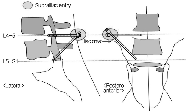
For the surgery on L5-S1 herniated disc, conventional posterolateral approach was performed instead of extreme lateral approach as iliac crest and large transverse process of L5 blocked access to L5-S1 disc space by trajectory of extreme lateral approach. Usually iliac crest is considered a major obstacle. Therefore, during L5-S1 level endoscopic discectomy, interlaminar approach instead of the posterolateral approach has been used for the endoscopic lumbar discectomy5,21). However, not all of the posterolateral approach is impossible for L5-S1 disc level. Yeung and Tsou25) performed 307 cases of endoscopic discectomy, about half of the cases were L5-S1 level discectomy and they performed all of them using posterolateral approach and reported successful post-operative prognosis in more than 81.4% of the cases. They reported that posterolateral approach can be used in any disc level of endoscopic lumbar discectomy. Compared to the interlaminar approach, the posterolateral approach is advantageous in removal of central type or extraforaminal herniated disc. Also during revision surgery of the patient treated with conventional microdiscectomy, the endoscopic discectomy with posterolateral approach for L5-S1 disc herniation is beneficial compared to the interlaminar approach as posterolateral trajectory passes unscarred virgin tissue and minimize iatrogenic neural injury5). When posterolateral endoscopic L5-S1 discectomy is performed, suprailiac entry is used due to the fact that the iliac crest conceals the L5-S1 disc space. As the skin entry point is cranial to the iliac crest, the trajectory is naturally angled to craniocaudal side (Fig. 6). There are cases where endoscopic approach is impossible due to unusually big height of the iliac crest or larger transverse process of L5. In this study, however, posterolateral approach to L5-S1 discectomy was possible in most cases, and if it was difficult, foraminoplasty was performed to approach disc space, the shaving off of ventral side of the superior facet of L55). In the research analyzing the relationship between L5-S1 disc and iliac crest and endoscopic discectomy of L5-S1, they report that if the height of iliac crest is higher than mid L5 pedicle, foraminioplasty is necessary to use transforaminal approach during the L5-S1 discectomy6).
Based on this study, complete removal of herniated disc was difficult, which decrease success rate of removal of severely migrated herniated disc. Lower limit of the endoscope is less than the half of the height of the lower vertebrae of the lesion, and when the herniated disc migrated to lateral recess, removal of the herniated disc was difficult due to the angle of approach was limited. When the herniated disc migrated to lateral recess, it could be approached from the different side, so this method should be considered. Or by making skin incision point more dorsal than usual and reach upper intervertebral foramen (intervertebral foramen consist of the upper vertebrae of the lesion) to increase the lower range of the endoscope, but increases nerve root damage as the exiting nerve root comes out of the upper intervertebral foramen. Limitations of this study are that the subject was specialized group of people and the long-term follow up was impossible. In addition, the result of the surgery is largely dependent on the proficiency of surgeon, but this research used data from three different surgeons’ cases. Despite all the limitations, this research provided insights for usability, stability, and limitation of the extreme lateral endoscopy compared to the previous endoscopic approach.
CONCLUSION
As young patients have soft intervertebral disc protrusion and less degenerative change, ELELD would replace the discectomy through laminectomy, but if the herniated disc migrates higher than the lower vertebral height in either upward or downward, or sequestrated, the removal of the herniated disc is extremely difficult, so the discectomy through laminectomy may be considered. As osteophyte due to the apophyseal ring fracture occurs frequently to the young patients, pre-operational CT scan must be performed and the discectomy through laminectomy or posterior lumbar interbody fusion is recommended.


