INTRODUCTION
Spinal epidural hematoma (SEH) is a rare neurological condition that occurs spontaneously or as a result of trauma. SEH can cause various neurological symptoms, which range from axial pain to severe neurological deficit, and may occasionally require urgent surgical decompression. Cervical SEH is rarer than other forms of spinal injuries. Without neurological deterioration, the diagnosis of SEH may be challenging and take a long time. We report a case of a patient who presented with post-traumatic cervical SEH accompanied with occipital neuralgia that was managed with conservative treatment.
CASE REPORT
A 32-year-old woman presented to our outpatient department with occipital headache. She had sustained injury during her swimming exercise a week earlier when the vertex region of her head had impacted the wall of a swimming pool. She had not lost consciousness and felt neither weakness nor sensory changes in her extremities at the time of injury. The day following the trauma, she developed an occipital headache that persisted for >1 week, so she underwent a brain computed tomography (CT) scan at a local clinic.
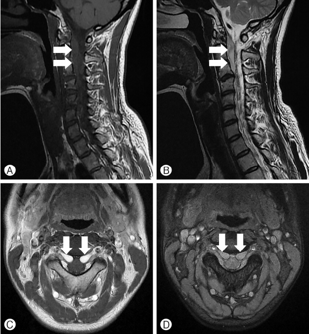
There was no abnormal finding from the CT scan, so she visited our outpatient department. She complained of a headache that persisted except during sleep. The occipital headache was bilateral and aggravated by flexion of the cervical spine. There was no external wound in her head and neck. Neurological examination revealed paresthesia of the occipital region. She exhibited no motor weakness. Plain X-ray imaging showed normal cervical spine alignment; furthermore, no fracture or dislocation was noted. Magnetic resonance imaging (MRI) findings revealed an epidural hematoma at the C1 to C3 level that was compressing the bilateral C2 roots (Fig. 1). No intramedullary lesion of the cervical spine was observed on MRI scan.
Surgical removal of the hematoma with decompression was not planned because there was no myelopathy or cord compression. A non-steroidal anti-inflammatory drug and gabapentin were prescribed for reducing the occipital neuralgia. A soft neck collar was applied to restrict cervical spine motion. Her symptoms gradually decreased and her occipital neuralgia eventually disappeared. One month after the trauma, MRI showed no residual epidural hematoma and she had no neurological deficits.
DISCUSSION
SEH is a very rare neurological condition. There are various causes of SEH that include vertebral fracture, lumbar puncture, post-surgical complications, epidural anesthesia, and anticoagulation therapy3,6,10,11). Unknown causes of SEH have been reported as spontaneous SEH, and a case of traumatic SEH has been recently reported. Previously, traumatic SEH has accounted for 0.5% to 1.7% of all spinal injuries1).
Symptoms of SEH vary according to the location of SEH along the spine. Compression of the root occurs with radiculopathy along the dermatome. Cord compression causes myelopathy and cord syndromes. Because of the variety of symptoms, SEH is sometimes not suspected at initial presentation and can reportedly occasionally mimic stroke or cord injury2,4,5,8,9). In polytraumatic patients, other injuries may mask SEH. Lin et al.7) reported that SEH was misdiagnosed as brachial plexus injury in a case of combined clavicle fracture. Careful examination and awareness of the possibility of SEH is required during differential diagnosis, and performing early MRI can yield useful results. In the present case, trauma was close to the cervical spine, and the presence of aggravated occipital neuralgia caused by cervical spine motion was evidently led to the diagnosis of SEH.
Surgical treatment is the gold standard for treating SEH showing acute severe neurological deterioration. However, several cases of spontaneous recovery of neurological deterioration without surgery have been reported. Some patients have improved during preparation for surgery. Rapid absorption also has been reported. Yi et al.12) reported resolution within three days after trauma, and the authors suggested epidural fat tissue or leakage into the paraspinal space via a neural foramen. Surgical treatment should be considered in patients with severe neurological deficits. During conservative management, symptomatic treatment and resting with restriction of spinal motion may be helpful.
SEH can cause various symptoms, and evaluation should be performed if SEH is suspected. Surgical and conservative treatment may be considered depending on the neurological symptoms of the patient.