INTRODUCTION
Schwannomas, also known as neurilemmomas and axonal intraneural Schwann cell tumors, are benign neoplasms of the nerve sheath originating from Schwann cells6). The most common schwannoma is acoustic neuroma. Schwannomas arise from the nerve sheath of large peripheral or cranial nerves and occur at the subcutaneous tissue level or in the deeper layers2). These tumors can arise from any nerve covered with a Schwann cell sheath, which includes the cranial nerves (except for the optic and olfactory nerves), spinal nerve, and autonomic nervous system7).
Ancient schwannomas are rare benign encapsulated tumors of protracted indolent growth; “ancient schwannoma” denotes extracranial schwannomas, which are usually solitary and grow to a large size. The term may also describe schwannomas that underwent pronounced cystic changes (e.g., relative loss of Antoni type A, perivascular hyalinization, calcification, and cystic necrosis). Cystic growth over the scalp can be easily misidentified as common tumors, such as dermoid cysts and trichilemmal cysts.
We present a rare case of an asymptomatic mass over the scalp; excision biopsy revealed this to be an ancient schwannoma.
CASE REPORT
A 36-year-old male patient presented to the department of neurosurgery at our institute complaining of a 3 × 2-cm palpable mass on the parietal area of the scalp that grew slowly over 5 years. The patient did not take any medications and had no relevant familial history.
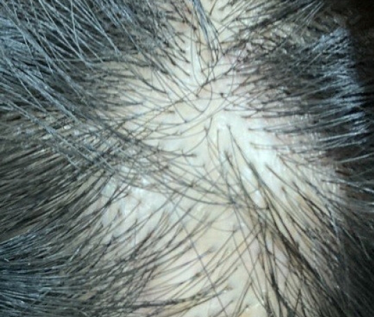
Physical examination revealed a solitary, round mass in the left parietal area of the scalp, which was soft and non-tender (Fig. 1).
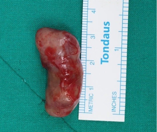
On February 3, 2022, we excised the mass under local anesthesia. Gross examination of the specimen revealed a well-encapsulated mass measuring 3 × 1-cm, with a grayish surface (Fig. 2).
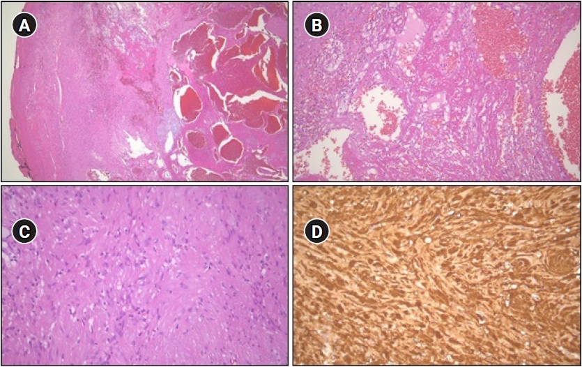
Thereafter, the specimen was sent for histopathological examination, which revealed a well-circumscribed mass with degenerative and cystic changes, as well as hemorrhage. The tumor cells were spindle-shaped without cytologic atypia and were positive for S-100 on immunostaining (Fig. 3). The histological features and immunohistochemical results were indicative of ancient schwannoma.
DISCUSSION
Schwannomas are among the most common benign nerve sheath tumors. However, the development of schwannoma on the scalp is rare and is easily mistaken for a tumor of hair. Especially trichilemmal cysts are common over the scalp and present as dermal or subcutaneous growths over scalp5).
Radiographic features of ancient schwannoma are calcification, hyalinization, and cystic cavitation.
Differed from ancient schwannoma, radiographic feature of trichilemmal cysts is nodules are small, ovoid, circumscribed subcutaneous masses without overlying skin ulceration or underlying bone erosion, and dermoid cysts which radiographically featured do not enhance after contrast administration.
Schwannomas can be divided into subtypes based on histological findings, such as ancient, microcystic/reticular, epithelioid, cellular, psammomatous, and melanotic schwannomas4). Ancient schwannoma is a rare subtype; it was first described by Ackerman and Taylor in 19511) and is characterized by increased cellularity and cystic necrosis. Additional histological characteristics include areas of diffuse fibrosis and hyalinization with nuclear pleomorphism and hyperchromasia but without nuclear atypia.
Our patient presented with an asymptomatic growth on the scalp for 5 years, confirmed to be an “ancient schwannoma,” different from a trichilemmal or dermoid cyst.
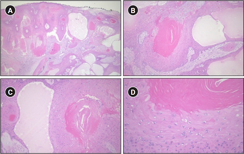
Trichilemmal cysts, common among middle-aged individuals, are smooth, mobile, and filled with keratin, a protein component found in hair, nails, skin, and horns. Histopathologically, trichilemmal cysts are lined by epidermal cells and contain homogeneous keratinous material (Fig. 4)3).
Dermoid cysts that develop from epithelium are often asymptomatic and are soft or rubbery, round, and subcutaneous nodule5).
When we first identified the patient’s lesion, trichilemmal cyst or dermoid cyst were the possible diagnoses. This was because these are commonly noted; thus, surgical treatment was accordingly planned. However, the result of immunohistochemistry revealed the tumor to be an ancient schwannoma, contrary to our expectations.
Although there was no major difference in the direction of treatment, the fact that histological results pointing to an uncommon disease are a possibility should be remembered.