| Home | E-Submission | Sitemap | Contact Us |  
top_img
The Nerve > Volume 3(2); 2017 > Article
Oh and Kim: Posterior Coccygeoplasty for a Presacral Tumor

Abstract

Presacral tumor is an uncommon tumor and it has potential for malignant transformation. There are various surgical approaches in presacral tumor surgery. It depends on the size and position of tumor and if it has invaded surrounding organs. Posterior coccygeoplasty has several beneficial aspects for a patient with presacral tumor. It is easy to access to lower lying presacral mass and effective to maintain functions of coccyx. Coccygeoplasty can prevent surgery related injuries of anal sphincter and sacrum. We present a 24-year-old female patient that underwent resection of a presacral tumor through posterior coccygeoplasty and review pertinent literature.

INTRODUCTION

The presacral tumor is an uncommon tumor and it has potential malignant transformation8,18). Diagnosis is usually delayed because it presents clinical symptoms raised from affecting structures by compression of tumor mass in late period of tumor growth5,7).
Posterior approach is preferred for small, benign lesions that do not extend above the level of the S2 vertebrae. The posterior approach is comprised of many techniques including the transsphincteric, transsacral, transrectal, transanorectal, and transsacrococcygeal approaches. Transcoccygeal approach only maintains anal sphincter and sacrum2). It is easy to access to lower lying presacral mass. Coccygeoplasty can maintain functions of coccyx and prevent surgery related injuries of anal sphincter and sacrum.
We present a 24-year-old female patient that underwent resection of a presacral tumor through posterior coccygeoplasty and review pertinent literature.

CASE REPORT

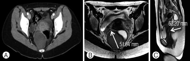
A 24-year-old woman had been treated with medication because of acute pyelonephritis for 3 weeks without improvement. She underwent computed tomography (CT) scan and magnetic resonance imaging (MRI) for further evaluation that revealed a 6.3cm-sized high density non-enhancing cystic lesion in the presacral area. Ovaries and uterus were intact (Fig. 1). There was no evidence of adenopathy or metastatic disease within the abdominal cavity. Physical examination revealed normal sphincter tone, normal flexibility of perianal soft tissue, and an empty rectal vault with extrarectal fullness posteriorly reachable with the fingertip.
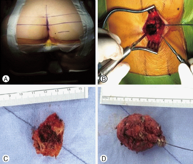
Posterior approach was conducted for total resection of tumor. Patient was placed in the prone jack-knife position with the buttocks spread (Fig. 2A). After an incision over the lower portion of the sacrum and coccyx down to the anus was conducted, the anococcygeal ligament was dissected and levator ani muscles were laterally retracted to obtain optimal surgical window approach to the presacral space (Fig. 2B). To provide good exposure, excision of the coccyx (coccygectomy) was necessary (Fig. 2C). Coccygotomy was conducted in a middle portion of coccyx because a part of bony coccyx should be retained for reconstruction of coccyx. Careful dissection was conducted to separate the tumor from the rectum and grossly total resection of tumor was conducted successfully (Fig. 2D). Reconstruction of coccyx was conducted using mini-profiled locking plates and monocortical (6mm length; 2.4mm diameter) locking screws (ARCH DePuy Synthes, Raynham, MA, USA). Bone dusts were packed into the bony cap between coccygectomy bone flap and remained coccyx. Wound closure was completed after irrigation and meticulous hemostasis.
Pathologic diagnosis was confirmed as an epidermoid cyst (Fig. 3). Adequate reconstruction of the coccyx was revealed on postoperative 1-year follow-up X-ray image (Fig. 4).

DISCUSSION

The presacral tumor is an uncommon tumor and may grow larger or become a malignant tumor8,13,20). About one-third of presacral tumors are congenital7,8,19); neurogenic and osseous are approximately 10% respectively, and etc.8). Approximately 10% of teratoma are transformed into malignancy in a few years6,8). Chordoma is most common solid retrorectal tumor3,4,7,8).
The method of diagnosis is varied. CT and MRI are effective in finding the size and position of tumor, relative to surrounding organs as well as other pertinent information. Biopsy is recommended for tissue confirmation despite possibility of post-procedure infection8). In addition, it may be routes of proliferation of malignant tumor cells.
Presacral tumor occasionally reveals non-specific symptoms3-5,8) such as abdominal pain, bowel habit change, constipation, and inguinal area pain. This often makes a diagnosis complicated and may produce a wrong diagnosis8,10,12,17,20).
Surgical removal of presacral tumor is preferred for reducing infection, preventing malignant transformation, and making an accurate diagnosis15,21).
Anterior and posterior surgical approaches may be applied for surgery of presacral tumor17). Selection of surgical approach depends on the size and position of tumor and if it has invaded surrounding organs12). The abdominal approach is usually conducted for these lesions that have their lowest extent above the level of the S4 vertebrae and do not have evidence of sacral involvement. Anterior approach is suitable for the tumor of upper sacral promontory20). Laparoscopic approach to removing these tumors is a safe and feasible for a part of presacral tumors1,11).
Posterior approach is preferred and familial for small, benign lesions that do not extend above the level of the S2 vertebrae2). The posterior approach is comprised of many techniques including the transsphincteric, transsacral, transrectal, transanorectal, and transsacrococcygeal approaches. Transcoccygeal approach only maintains anal sphincter and sacrum2). It is easy to access to lower lying presacral mass. But, transcoccygeal approach needs resection of coccyx. Coccygeoplasty can maintain functions of coccyx and prevent surgery related injuries of anal sphincter and sacrum.
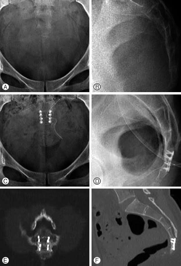
Coccygotomy should be conducted in a middle portion of coccyx and a part of bony coccyx should be retained. Bone on bone fusion should be conducted for complete reconstruction of coccyx and final X-ray revealed stable reconstruction and bony fusion for this patient (Fig. 4).
Transsphincteric approach may cause sphincter deterioration9,14,16) and transsacral approach may cause hip discomfort and fistula11,20). Even though posterior approaches cannot be entirely excluded from surgery related complications, coccygeoplasty has merits such as presacral organ protection, stability in sacrococcygeal area, and stable sitting position.

CONCLUSION

Posterior coccygeoplasty may be effective in maintaining bony structure, anal sphincter function, and preservation of coccyx for patients with lower lying presacral mass.

CONFLICTS OF INTEREST

No potential conflict of interest relevant to this article was reported.

Fig. 1
(A) A 6.3 cm non-lobulated cyst with moderate signal intensity was shown on axial view of computed tomography scan image. Cystic mass was no definite enhancing portion on (B) axial and (C) sagittal views of abdomen/pelvic magnetic resonance image.
nv-3-2-78f1.gif
Fig. 2
(A) The operation was conducted in prone jack-knife position. (B) Incision was made on the lower portion of the sacrum and coccyx and (C) coccyx was removed as a one piece after coccygotomy. (D) Total resection of tumor was conducted.
nv-3-2-78f2.gif
Fig. 3
Squamous epithelium (A) and lamellated keratin (B) were shown on the specimen slide image. Cystic cavity filled with lamellated keratin lined by a squamous epithelium (H & E, ×200).
nv-3-2-78f3.gif
Fig. 4
(A, B) Preoperative and (C, D) postoperative sacrococcygeal X-rays are shown. (E, F) Solid bone fusion was confirmed on computed tomographic scan 1 year later after surgery.
nv-3-2-78f4.gif

REFERENCES

1. Bax NM, van der Zee DC: The laparoscopic approach to sacrococcygeal teratomas. Surg Endosc 18:128-130, 2004
crossref pmid
2. Canessa CE: Dorsal transsacrococcygeal rectal approach. Dis Colon Rectum 48:1663-1665, 2005
crossref pmid
3. Cody HS 3rd, Marcove RC, Quan SH: Malignant retrorectal tumors: 28 years’ experience at Memorial Sloan-Kettering Cancer Center. Dis Colon Rectum 24:501-506, 1981
crossref pmid
4. Freier DT, Stanley JC, Thompson NW: Retrorectal tumors in adults. Surg Gynecol Obstet 132:681-686, 1971
pmid
5. Glasgow SC, Birnbaum EH, Lowney JK, Fleshman JW, Kodner IJ, Mutch DG, et al.: Retrorectal tumors: a diagnostic and therapeutic challenge. Dis Colon Rectum 48:1581-1587, 2005
crossref pmid
6. Head HD, Gerstein JD, Muir RW: Presacral teratoma in the adult. Am Surg 41:240-248, 1975
pmid
7. Hobson KG, Ghaemmaghami V, Roe JP, Goodnight JE, Khatri VP: Tumors of the retrorectal space. Dis Colon Rectum 48:1964-1974, 2005
crossref pmid
8. Jao SW, Beart RW Jr, Spencer RJ, Reiman HM, Ilstrup DM: Retrorectal tumors. Mayo Clinic experience, 1960-1979. Dis Colon Rectum 28:644-652, 1985
crossref pmid
9. Kanemitsu T, Kojima T, Yamamoto S, Koike A, Takeshige K, Naruse T: The trans-sphincteric and trans-sacral approaches for the surgical excision of rectal and presacral lesions. Surg Today 23:860-866, 1993
crossref pmid
10. Kolodziejski LS, Dyczek ST, Pogodzinski M: Surgical management of retrorectal tumors. J Chir (Paris) 141:109-113, 2004
pmid
11. Konstantinidis K, Theodoropoulos GE, Sambalis G, Georgiou M, Vorias M, Anastassakou K, et al.: Laparoscopic resection of presacral schwannomas. Surg Laparosc Endosc Percutan Tech 15:302-304, 2005
crossref pmid
12. Lev-Chelouche D, Gutman M, Goldman G, Even-Sapir E, Meller I, Issakov J, et al.: Presacral tumors: a practical classification and treatment of a unique and heterogeneous group of diseases. Surgery 133:473-478, 2003
crossref pmid
13. Lovelady SB, Dockerty MB: Extragenital pelvic tumors in women. Am J Obstet Gynecol 58:215-236, 1949
crossref pmid
14. Meissner K, Jirikowski B, Szecsi T: Mason paracoccygeal transsphincteric approach to the rectum: good outcome only in highly selective indications? Chirurg 67:145-149, 1996
pmid
15. Pappalardo G, Frattaroli FM, Casciani E, Moles N, Mascagni D, Spoletini D, et al.: Retrorectal tumors: the choice of surgical approach based on a new classification. Am Surg 75:240-248, 2009
crossref pmid pdf
16. Pescatori M, Brusciano L, Binda GA, Serventi A: A novel approach for perirectal tumours: the perianal intersphincteric excision. Int J Colorectal Dis 20:72-75, 2005
crossref pmid
17. Singer MA, Cintron JR, Martz JE, Schoetz DJ, Abcarian H: Retrorectal cyst: a rare tumor frequently misdiagnosed. J Am Coll Surg 196:880-886, 2003
crossref pmid
18. Spencer RJ, Jackman RJ: Surgical management of precoccygeal cysts. Surg Gynecol Obstet 115:449-452, 1962
pmid
19. Uhlig BE, Johnson RL: Presacral tumors and cysts in adults. Dis Colon Rectum 18:581-589, 1975
crossref pmid
20. Wolpert A, Beer-Gabel M, Lifschitz O, Zbar AP: The management of presacral masses in the adult. Tech Coloproctol 6:43-49, 2002
crossref pmid
21. Woodfield JC, Chalmers AG, Phillips N, Sagar PM: Algorithms for the surgical management of retrorectal tumours. Br J Surg 95:214-221, 2008
crossref pmid
TOOLS
PDF Links  PDF Links
PubReader  PubReader
ePub Link  ePub Link
XML Download  XML Download
Full text via DOI  Full text via DOI
Download Citation  Download Citation
  Print
Share:      
METRICS
1
Crossref
11,994
View
108
Download
Related article
Editorial Office
Department of Neurosurgery, Dankook University Hospital
201, Manghyang-ro, Dongnam-gu, Cheonan-si, Chungcheongnam-do 31116, South Korea
TEL: +0082-41-550-6280   FAX: +82-41-556-0524   
E-mail: spine1225@naver.com , mousehunt7@naver.com
About |  Browse Articles |  Current Issue |  For Authors and Reviewers
Copyright © The Korean Society of Peripheral Nervous System.                 Developed in M2PI