INTRODUCTION
Patients presenting with scalp mass are frequently encountered in daily practice4). The spectrum of causes is comprehensive. They might be directly related to the scalp itself. They might be secondary stigmata of an underlying process in the skull. The overall incidence of specific diagnosis and rates of clinically significant lesions presenting as scalp masses have not been well-defined4). According to a study to determine the incidence of scalp mass in adults4), the five most common histological diagnosis are trichilemmal cyst, epidermal cyst, lipoma, nevus, and sebaceous cyst. The rate of “clinically significant” pathologies meaning malignancies or those that need follow-up is approximately 7.8%4). Scalp masses originated from neural components included neurofibroma and Merkel cell carcinoma, although their incidence is quite low (0.6 and 0.3%, respectively)4). Preoperative diagnosis of scalp mass is difficult4).
Benign peripheral nerve sheath tumors (PNSTs) such as neurofibromas and schwannomas may present as extracranial head and neck mass3). However, their presentation as small and tender scalp mass is extremely rare. Here, we report two cases of PNSTs presented as small and tender scalp masses.
CASE REPORTS
1. Case 1
A 56-year-old female presented a small and palpable tenderness on her left temporal area. Until feeling electricity-like pain and tenderness in her left temporal area, she did not realize the presence of this lesion 2 months before presentation. There was no spontaneous pain or headache. A brief episode of electricity-like pain was elicited when touching the mass. It was movable, showing rubbery in consistency. Her medical and family history was unremarkable. Her neurologic examination was normal. There was no hypesthesia or paresthesia around the mass. No abnormal skin pigmentation or café au lait spot was observed. An excision biopsy was performed and a 3×8mm-sized whitish mass was excised. Histologic examination revealed a neurofibroma (Fig. 1). No recurrence was observed with 12 months of follow-up.
2. Case 2
A 56-year-old female presented a small tenderness in her right retroauricular occipital area for three months. She realized the presence of a small palpable mass in her occipital scalp one year prior to presentation. It showed gradual enlargement. There was no pain or tenderness at the beginning. However, aching pain and tenderness gradually developed. At one and a half years prior to presentation, she underwent an excisional biopsy for a small tender scalp mass adjacent to the location of the present one. At that time, no histologic examination was performed. Her postoperative course was uneventful. Due to increasing pain intensity and progressive enlargement of her occipital mass, she was referred for histologic diagnosis.
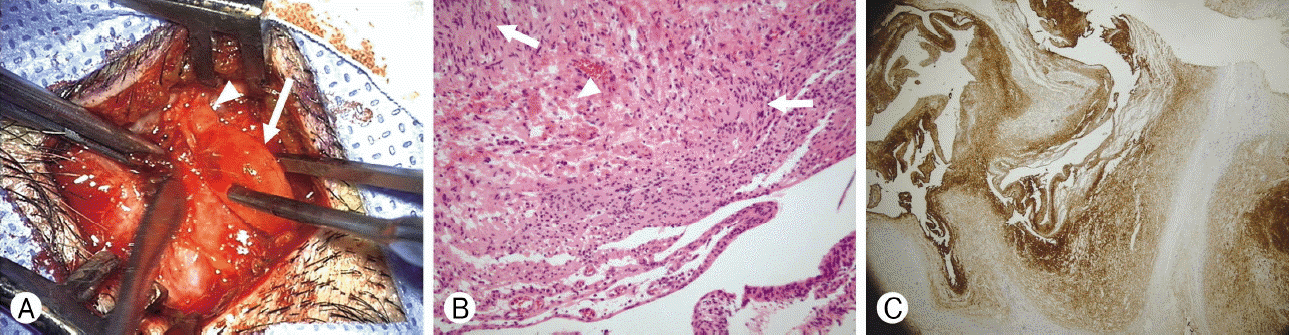
Her neurologic examination was normal. Her family and medical history were unremarkable. Manipulation of the mass elicited an incident and sharp pain over the mass with radiating paresthesia over the parieto-occital scalp. There was no café au lait spot or other skin stigmata of neurofibromatosis. An excisional biopsy revealed a 4×10mm-sized, fusiform swelling of a small branch of the lesser occipital nerve (Fig. 2A). The mass was excised en-bloc. Histologic examination revealed a schwannoma. Immunostaining for S-100 was positive. Neuro-filament was negative with a localized focus of increased Ki-67 (up to 15% was noted within the mass). Mild paresthesia was observed in her parieto-occipital scalp at 3 months postoperatively. There was no recurrence or new occurrence of scalp mass at the last follow-up at postoperative 12-month.
DISCUSSION
Differential diagnosis and management strategies for scalp masses have not been described in details yet. Their clinical significance also remains unclear. It is difficult to refer to the literature regarding the overall incidence of particular diagnosis and rates of clinically significant lesions presenting as scalp masses4). Scalp masses have been underestimated. Interdisciplinary consultations have not been performed for scalp masses comprehensively4). According to a recent study in a single center4), the overall incidence of clinically significant pathologies of scalp mass is around 7.8%. The incidence of correct preoperative diagnosis for scalp mass is only 13 to 27%4). Preoperative diagnosis of scalp mass is difficult4). It is not specified by the surgeon in 53% of patients. Even if it is specified preoperatively, preoperative diagnosis is correct in just 13 to 27% of these cases with respect to surgical specialties4).
In the report of Türk et al.4), for 345 patients with scalp masses, the most common pathology is found to be trichilemmal cyst, followed by epidermal cyst, lipoma, nevus, and sebaceous cyst. There five diagnoses account for 77.1% of these cases4). Tumors originating from cutaneous appendages (trichilemmal cyst, pilomatrixoma, sebaceous cyst, sebaceous carcinoma, apocrine mixed tumor, syringocystadenoma, hidradenoma, eccrine cell carcinoma, and nevus sebaceous) have constituted almost half of these cases (49%)4). A total of 82.3% of these lesions have originated from skin components, the epidermis, or cutaneous appendages4). The rest lesions are tumors of fat tissue, muscles, cartilage, bones, neural and neuroendocrine components, vascular tissue, proliferation of fibrous or related tissues, and cutaneous metastases or cutaneous lymphoid infiltrations. In their series, there were 2 (0.6%) cases of neurofibroma without any case of schwannoma. Although huge scalp masses due to PNSTs have been sporadically reported, their presentation as small and tender scalp mass is rare.
Among benign tumors of peripheral nerves, one specific group originated from Schwann cells is currently divided into two subtypes: neurofibromas and schwannomas3). Schwannomas also arise from encapsulated masses on the periphery of nerves. They contain characteristic cellular and spindled Antoni A areas with distinctive nuclear palisading (Verocay bodies) and looser Antoni B areas2). Entrapped axons are not their common features. In contrast, neurofibromas will diffusely expand the involved nerve and encompass entrapped axons. Schwannomas commonly arise from intracranial nerves while neurofibromas rarely affect cranial nerves. Neurofibromas may undergo malignant degeneration while schwannomas very rarely do so5).
Schwannomas occurring in the head and neck region account for approximately 25% of these cases. They are sometimes associated with von Recklinghausen’s disease in 8 to 18% of cases. When cases are not associated with neurofibromatosis as depicted in the current report, schwannomas are clinically seen as solitary slow-developing lesions with symptoms only when large areas have been affected1). Presentation might be pain along the distribution of nerve, hypesthesia and tenderness. Extradural schwannomas are most commonly found in association with large nerve trunks where motor and sensory modalities are intermixed2).