Tarsal Tunnel Syndrome: A Narrative Review
Article information
Abstract
Tarsal tunnel syndrome is a compressive neuropathy of the posterior tibial nerve beneath the flexor retinaculum on the medial ankle. Several intrinsic or extrinsic factors may contribute to pain over the medial plantar aspect of the foot that is aggravated by activities. In the presence of suggestive clinical features, appropriate use of radiographic and electrodiagnostic tests can be helpful. Surgical management is considered in patients who do not respond to non-operative treatment or those who have a space-occupying lesion within the tarsal tunnel. Along with surgical decompression of the posterior tibial nerve, complete removal of the causative space-occupying lesion is needed to prevent recurrence.
INTRODUCTION
Tarsal tunnel syndrome is defined as an entrapment neuropathy of the tibial nerve within the tarsal tunnel, which is a fibro-osseous tunnel beneath the flexor retinaculum on the medial ankle1). It was first described by Kopell and Thompson in 196011) and was named “tarsal tunnel syndrome” by Lam in 196212). The condition is similar to the carpal tunnel syndrome of the wrist concerning their analogous nerve compressive etiology. However, tarsal tunnel syndrome occurs less frequently and is often underdiagnosed or misdiagnosed. In addition, its therapeutic strategy should be established in a different perspective compared with other compressive neuropathies, especially in surgical decompression5). In this narrative review, we attempted to introduce the relevant anatomy, overall etiology, diagnosis, and management of this nerve entrapment syndrome.
ANATOMY
The tarsal tunnel is a fibro-osseous space located behind the medial malleolus. Its floor is formed by the medial wall of the talus, calcaneus, and distal tibia. It is covered by the flexor retinaculum, which attaches from anteromedial surface of medial malleolus and runs posteriorly onto the medial tuberosity of calcaneus. The tunnel is occupied by the tendons (tibialis posterior, flexor digitorum longus, and flexor hallucis longus) and the neurovascular structures (the posterior artery and veins and the posterior tibial nerve)2) (Fig. 1). The posterior tibial nerve, which is a branch of the sciatic nerve, runs down the back of the leg and enters the tunnel proximally, and in 93% of cases, the nerve branches into its three terminal branches: medial plantar nerve, lateral plantar nerve, and medial calcaneal nerve within the tunnel7). The medial and lateral plantar nerves supply autonomic, sensory, and motor fibers to the plantar foot. The medial calcaneal nerve passes through the flexor retinaculum and provides sensory innervation to the heel5). In 25% of cases, there is a variation where it may branch before and run superficial to the flexor retinaculum or arise from the lateral plantar nerve1).
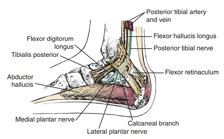
Schematic diagram of the anatomy of the tarsal tunnel.
ETIOLOGY
Several intrinsic or extrinsic factors may bring about compression of the posterior tibial nerve and its branches. The intrinsic factors include hypertrophic retinaculum, tendinopathy, arterial insufficiency, or space-occupying lesions within the tarsal tunnel, such as ganglion cysts, perineural fibrosis, osteophytes, calcaneal osteochondroma, lipomas or other tumors (Fig. 2-4). Extrinsic factors can be sequelae of ankle trauma, foot deformities, poorly fitted shoes, overweight, or systemic disease (Fig. 5, 6). It is also known to be more prevalent in runners, especially when they experience hyper-pronation of the foot8). In 20% of cases, however, the etiology of tarsal tunnel syndrome remains unclear and is considered idiopathic.

Ganglion cyst. A 23-year-old woman presenting with pain and paresthesia on the medial plantar aspect of the foot. (A, B) Fat-suppressed proton density magnetic resonance images show a high signal intensity mass (arrows) located around the neurovascular bundle (blank arrow). (C) Intraoperative image showing a ganglion cyst (asterisk) within the tarsal tunnel.
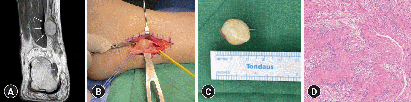
Schwannoma of the posterior tibial nerve. A 62-year-old man presenting with a painful, palpable mass over the medial ankle. (A) A T2-weighted magnetic resonance image shows a well-defined lesion with high signal intensity arising from the posterior tibial nerve. (B) Intraoperative finding of a mass arising from the posterior tibial nerve. (C) A well-circumscribed, encapsulated solid mass revealed a diffusely myxoid and homogenously grayish-yellow cut surface. (D) Histological features exhibit neurogenic, spindle cell proliferation with biphasic pattern of growth and prominent nuclear palisading (hematoxylin and eosin stain, ×100 magnification).

Osteochondroma of the calcaneus. A 19-year-old man presenting with insidious onset of burning pain accompanied by paresthesia and a palpable mass on the left foot. (A) A computed tomography image shows a 2.5 × 1 cm bony protuberance below the sustentaculum tali, demonstrating an apparent corticomedullary continuity with the underlying bone (arrow). (B) A T2-weighted fat-saturated magnetic resonance image shows a high signal intensity mass surrounded by a thin cartilaginous cap, which extends from the sustentaculum tali (arrow).

Calcaneal open fracture. A 65-year-old man presenting with burning pain and open wound over the medial ankle after a traffic accident. (A) Computed tomography image shows a calcaneus fracture with a displaced medial sustentaculum tali fragment (arrow). (B) Open wound on the medial side of the ankle. (C) After open reduction of the fracture, the posterior tibial nerve (arrow) was decompressed.

Valgus hindfoot alignment. A 59-year-old woman diagnosed with tarsal tunnel syndrome on the right foot showed heel valgus deformity (dotted circle).
DIAGNOSIS
The typical patient complaint is pain around the tarsal tunnel, with irradiation to the planar aspect of the foot. A sensation of tightness and to varying degrees sensory symptoms of burning, tingling and numbness are usually present. Symptoms worsen with weight-bearing activities such as prolonged standing or walking and are relieved by rest and leg elevation.
In the physical examination, neurological deficits are rare3). Mild sensory loss can sometimes be accompanied, although its precise assessment is difficult because of calluses on the sole of the foot. Deep palpation of the medial aspect of the ankle reveals tenderness with a positive Tinel’s sign. Provocation test which is performed by passive dorsiflexion and eversion of the foot while dorsiflexion the metatarsophalangeal joints may be helpful10).
Radiographic evaluation of the foot and ankle should be obtained to evaluate structural abnormalities such as tarsal coalitions, osteophytes, hindfoot varus/valgus, or evidence of previous trauma. If a space-occupying lesion is suspected, a diagnostic ultrasound or MRI scan can be obtained. MRI is known to provide 80% accuracy for identifying space-occupying lesions in tarsal tunnel syndrome and is considered the gold standard imaging technique9) (Fig. 7).

A diagnostic flow diagram for the assessment of tarsal tunnel syndrome. MRI: magnetic resonance imaging; USG: ultrasonography.
An electrodiagnostic study is helpful in differentiating the nerve compression within the tarsal tunnel from generalized neuropathy or radiculopathy15). However, its sensitivity and specificity remain low, and its results do not confirm tarsal tunnel syndrome13). In other words, with definite clinical symptoms and physical findings that correlate with tarsal tunnel syndrome and the failure of a patient to respond to conservative therapy, negative electrodiagnostic testing does not provide a contraindication for surgery.
MANAGEMENT
1. Non-operative
Tarsal tunnel syndrome is first treated non-operatively. Symptoms can be controlled by anti-inflammatory medication or orthotic devices that can modify biomechanical abnormalities and unload the tarsal tunnel. Activity modification combined with progressive mobilization exercise is encouraged14). Physiotherapy including taping, bracing, stretching, icing, massage, and ultrasound can also be used. Rarely, a local corticosteroid injection into the tarsal tunnel can be considered, with the potential risk of tendon rupture. Overall, non-operative approaches may in part be effective in relieving symptoms but lack sufficient evidence for their efficacy in complete remission of the nerve entrapment pathophysiology.
2. Operative
Surgical intervention for tarsal tunnel syndrome is indicated among symptomatic patients who do not respond to non-operative treatment or those who have space-occupying lesions within the tarsal tunnel. Because chronic nerve compression leads to intraneural fibrosis which can be responsible for the later development of muscle wasting and weakness, surgical decompression is recommended within 10 to 12 months from onset of symptoms17,18). In the literature, success rates following the surgery vary from 44% to 96%16). Among many factors that contribute to a satisfactory outcome after the surgery, a positive Tinel’s sign is considered one of the most predictable indicators4).
Tarsal tunnel release involves releasing the flexor retinaculum from its proximal attachment near the medial malleolus down to the sustentaculum tali and splitting the deep fascia of the abductor hallucis muscle to eliminate any possible compression of the terminal branches of the tibial nerve. The deep fascia of the abductor hallucis muscle is known to be the region where the greatest compression occurs but is often neglected and not addressed during the tarsal tunnel release1). Neurolysis is not typically performed because doing so can make the nerve more vulnerable to scarring by disrupting its surrounding bed and damaging the vaso nervosum. During the release, it is crucial to meticulously remove causative space-occupying lesion within the tarsal tunnel to prevent recurrence and residual symptoms6,19) (Fig. 8). At the end of the procedure, the tourniquet, if one was used, should be deflated. The nerve is then closely observed to determine whether capillary filling is adequate along its course. If the nerve does not “pink up” in certain areas, which can represent an area of constriction, release of the epineurium overlying the nerve may be needed. Any bleeding must be controlled, and the wound is closed without closing the retinaculum.
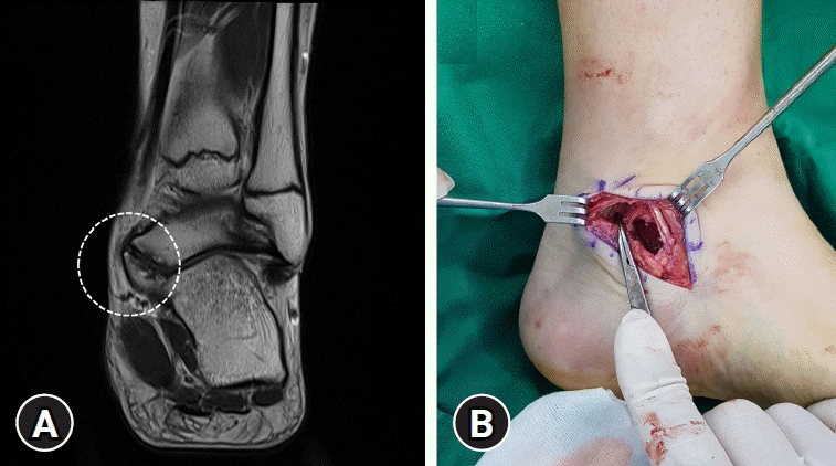
An 18-year-old man presenting with a painful mass over the medial ankle and decreased subtalar joint motion. (A) A T1-weighted magnetic resonance image shows a talocalcaneal coalition (dotted circle). (B) Sufficient void space was present around the posterior tibial nerve after resection of the coalition.
Recurrence rate after tarsal tunnel release varies from 5% to 60%. Possible reasons for the failed procedure are as follows: inaccurate diagnosis, inadequate release, insufficient hemostasis leading to scarring around the nerve, persistent nerve hypersensitivity, and pre-existing or iatrogenic nerve damage6). If every effort of conservative measures fails, revision surgery can be considered.
CONCLUSION
Tarsal tunnel syndrome is a significantly confusion condition and should be diagnosed based on a detailed history and clinical examination. In the presence of suggestive clinical features, appropriate use of radiographic and electrodiagnostic tests can be helpful. Nonoperative treatment can first be attempted but surgery is indicated among symptomatic patients who do not respond to non-operative treatment or those who have space-occupying lesions within the tarsal tunnel. Surgical intervention should be focused not only on complete decompression of the nerve but also on meticulous removal of causative space-occupying lesions to prevent recurrence.
Notes
No potential conflict of interest relevant to this article was reported.