Rubral Tremors Associated with an Inferior Olivary Lesion that Developed after a Brainstem Hemorrhage
Article information
Abstract
Rubral (Holmes’ or midbrain) tremor caused by a dentate projection pathway is a rare combination of 2 to 5 Hz rest, enhanced by posture and kinetic tremors of an upper extremity. Symptomatic palatal tremor is a defined movement disorder characterized as a 1–3 Hz palatal myoclonus that appears after a disruption of the Guillain-Mollaret triangle, which includes the ipsilateral red nucleus, the ipsilateral inferior olivary nucleus (ION), and the contralateral dentate nucleus. Although palatal tremor remains the only dyskinesia that is consistently related to hypertrophic olivary degeneration (HOD), some uncommon dyskinesias may occur with HOD. Both of these tremors can sometimes occur after a brainstem lesion. In this study, a 66-year-old female patient underwent a ventriculo-peritoneal shunt for hydrocephalus after a coiling ruptured paraclinoid aneurysm of the right side. At the time, the right brainstem and cerebellar hemorrhagic infarction had been identified. In a follow-up imaging, hemorrhagic infarction had improved, but the patient gradually developed a rubral tremor involving both the left upper and lower extremities, besides the head and tongue tremors. Rubral tremor occurred about three months after bleeding, and the head and tongue tremors about five months. Usually, rubral tremor is in the arm, but in this case symptoms appeared even in the legs, accompanied by head and tongue tremors. The literature and imaging findings of this uncommon condition are reviewed.
INTRODUCTION
In 1904, Gordon Holmes identified rest, postural, and action tremor movement disorders that occurred at less than 5 Hz. These types of tremors are named rubral, Holmes’ or midbrain tremor. Damage to the cerebellothalamic tract is thought to be involved in this tremor. Reported foci include the area superolateral to the red nuclei, rubrothalamic tract, central tegmental tract, superior cerebellar peduncle and the substantia nigra1,7). Rubral tremor occur as a consequence of a lesion in both the dopaminergic and the non-dopaminergic (cerebellothalamic/ cerebelloolivary) systems3).
Palatal tremor or myoclonus has been reported to be associated with hypertrophic olivary degeneration (HOD). The tremor is known to cause damage to Guillain-Mollaret triangle - the dentate-rubro-olivary tract. Palatal contractions are seen at a frequency of 1 to 3 Hz. HOD usually occurs unilaterally and ipsilateral to the lesion if the lesion is in the brain stem, or contralateral to the lesion if the lesion is in the cerebellum, as identified in MR images4).
The initial MR showed that our patient had brainstem hematoma. After that, the lesion evolved unilateral HOD. The patient had rubral tremor on the upper and lower extremities followed by symptomatic palatal tremor.
CASE REPORT
The 66-year-old woman receiving treatment for hypertension was admitted to the emergency room with a thunderclap headache. Subarachnoid hemorrhage (SAH) due to a ruptured-paraclinoid aneurysm of the right side was observed by computed tomography (CT) (Fig. 1), and the patient immediately underwent coil embolization for the aneurysm. In addition, thick SAH was seen around the quadrigeminal cistern of the right side on CT. Moreover, extensive intracranial hemorrhage was also observed in the midbrain, pontine tegmentum and cerebellum on the right side. The neurologic examination revealed that the patient had a stuporous mentality and hemiparesis of the left side of motor grade II/V. Two months later, the patient underwent a ventriculo-peritoneal shunt due to persistent hydrocephalus. After 1 month, a right inferior olivary nucleus, and red and dentate nucleus lesions longitudinally appeared on an MRI scan (Fig. 2).
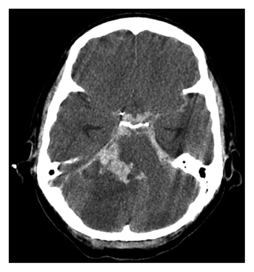
Pre-contrast brain CT image at 1 day after coil embolization. It showed thick SAH around interpeduncular, crural and quidrigeminal cisterns, com pressed brainstem and bilateral cerebellar infarction.

Hemorrhage and edema were seen on the MRI brain scan after 1 month, extending from the dorsal midbrain to the cerebellum.
In the intensive care unit, the patient gradually developed rubral tremor involving all extremities - more dominantly on the right side - and subsequent head and tongue tremors. In other words, rubral tremor occurred about three months after the bleeding, and head and tongue tremor about five months. The tremor in the extremities manifested as a slow, regular and low-amplitude tremor at rest, and an irregular, highamplitude and low-frequency (<4Hz) tremor enhanced by posture and movement. After 10 months, the lesion of the unilateral dorsal midbrain and dentate nucleus, HOD on the right side, were observed on MRI (Fig. 3). Also, on the EEG, intermittently slow and generalized waves were observed. The patient was prescribed antiepileptic drugs, such as levetiracetam, topiramate and phenytoin for abnormal EEG waves, and clonazepam and levodopa (Starlevo®) for tremors, but these drugs did not significantly improve the symptoms.
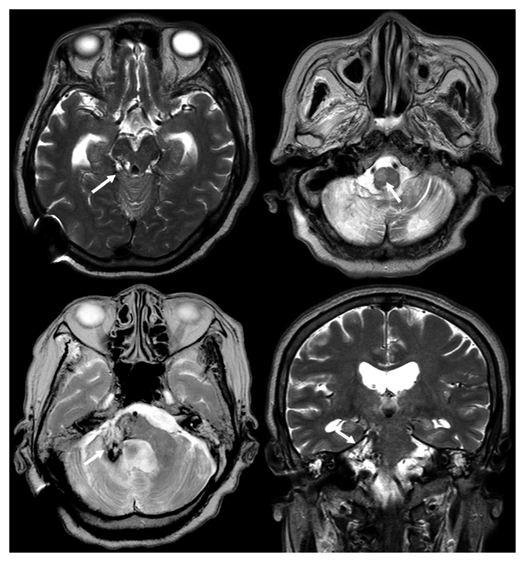
Dorsal midbrain lesion and unilateral HOD of the right side as seen on MRI brain scan after 10 months. This lesion could result in rubral tremor and symptomatic palatal tremor.
DISCUSSION
In this case, rubral tremor on the right side of the patient was more dominant despite the brainstem lesion being mainly on the right, but this is thought to be the result of weakness on the left side due to the initial hemorrhage. We suggest that the tremors on both sides could be the result of lesions of bilateral cerebellar hemorrhagic infarction and Guillian - Mollaret triangle lesions - ipsilateral central tegmental tract and dentate nucleus - due to hemorrhagic infarction extensively from the midbrain to the medulla. This triangle is an important network connecting the dentate nucleus of the cerebellum of one side with the red nucleus and the inferior olivary nucleus on the contralateral side, via the superior cerebellar peduncle, the central tegmental tract and the inferior cerebellar peduncle, respectively. There are numerous reports of such results4,9). The duration between the lesion and the first occurrence of the tremor is variable from 4 weeks to 2 years. Depending on the location of interruption, cerebellar atrophy or HOD can occur. The abnormality of the inferior olivary nucleus is thought to be the result of transsynaptic degeneration due to a lesion either in the ipsilateral central tegmental tract or in the contralateral dentate nucleus10). Holmes reported that if the tremor was due to a lesion of the cerebello-rubro-spinal system in any part of it, the tremor should be associated with a homolateral lesion of the superior cerebellar peduncle, or the origin of this is the nucleus dentatus of the cerebellum5). Another possibility is thought to be due to a lesion that had compressed the other side triangle through the midline. A similar case has been reported in some other studies with respect to the HOD4).
In our case, head and tongue tremors are also thought to be clinically associated with the right HOD on MRI. We considered carefully whether this tremor was associated with the palatal tremor. Unfortunately, we did not conduct any tests, such as ultrasound, to determine the cause. However, there have been reports that may support this possibility. Futaba Maki et al. reported a rubral tremor that was the result of the disruption of afferent pathways (deafferentation) from the cerebellar dentate nucleus to the thalamus due to a brainstem lesion. Meanwhile, palatal tremor is due to a disruption of the pathways from the dentate nucleus to the inferior olivary nucleus via the central tegmental tract, which result in HOD6). In this case, these lesions were identified. As in our case, there have been reported cases of rubral tremor associated with the lower extremities8). Our patient experienced the tremor, mainly on the right side of her body, most significantly below the ankle, which was also accompanied by a dystonic feature of the big toe. The exact anatomical reason was still not revealed, but the cerebellothalamic tract and dopaminergic nigrostriatal system are thought to be involved2,8).
As noted earlier, many studies have reported that the involvement of the cerebello-olivary system or cerebellar outflow tract in a bilateral disruption of the rubro-olivo-cerebellorubral loops may be a reasonable explanation for the significant findings in our patient, although this may not able to explain all her symptoms. To our knowledge, there are only a few reports to date of uncommon dyskinesia similar to rubral tremor with probable palatal tremor. In conclusion, our case suggests that the involvement of the bilateral cerebellar-olivary system and its outflow pathways may have led to two distinct tremors.
CONCLUSION
In this case, rubral tremor on the right side of the patient was more dominant despite the brainstem lesion being mainly on the right, but this is thought to be the result of weakness on the left side due to the initial hemorrhage. Both left and right side tremors had resulted from bilateral cerebellar and Guillian-Morallet triangle lesion due to hemorrhagic infarction extensively from the midbrain to the medulla. Another reason is thought to be due to the lesion that compressed the triangle on the other side through the midline. Moreover, head and tongue tremors are thought to be clinically associated with the right HOD on MRI. Our case showed that the involvement of the bilateral cerebellar-olivary system and its outflow pathways, may have led to two distinct tremors.